Virtual EyePath Slides
 |
 |
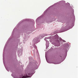
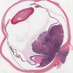
| Conjlymhoma | RBoptnerveinvasion |
 |
 |
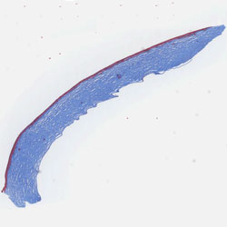
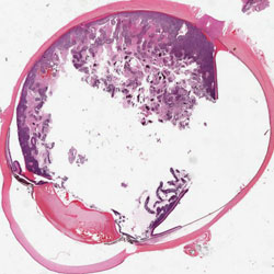
| Granular | Rbrosettes |
Virtual microscopy is a novel method of posting microscope images on, and transmitting them over, computer networks for the purpose of facilitating collaborative interaction among colleagues across diverse geographical locations. It involves a synthesis of microscopy technologies and digital technologies. With recent advances in virtual microscopy, it is now possible to achieve image resolutions approaching that visible under the optical microscope. View Site
An ophthalmic pathology virtual microscopy workgroup has been established in order to create an Ophthalmic Pathology Collaborative and Educational Resource (OPCER) for ophthalmologists, ophthalmology residents, and medical students. Additional potential benefits include Continuing Medical Education for ophthalmologists and eye pathologists, and Quality Assurance programs for practicing eye pathologists. The following institutions are part of this OPCER workgroup:
- Loyola University Medical Center
- Summa Health System
- Duke University
- University of Iowa, Carver College of Medicine
- Northwestern University, Feinberg School of Medicine
- The New York Eye and Ear Infirmary
- Rush University Medical Center
High quality histopathologic specimens for ophthalmic pathology are scanned using technology available from Aperio. We currently have a large data base of scanned specimens which are available for viewing. Specimens are annotated with educational information. Ultimately, the goal of this project is to provide an invaluable ophthalmic pathology resource which will be made available to interested individuals worldwide.